Biomedical Core Facility
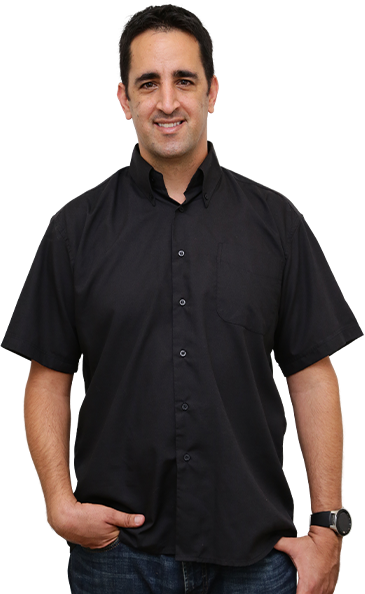
Ziv Itach Abramovitch
CORE FACILITY MANAGER
Online Registration System
Biomedical innovators’ one-stop resource for state-of-the-art technology and expertise. We are committed to provide outstanding resources to all researchers both in academia and industry. Our mission is to support biomedical research by providing accessibility to innovative and advanced scientific technologies. Our professional personnel assist the researchers during planning, execution and analysis of experiments.
Contact
Info
Contact Info
Primary Contact
Ziv Itach Abramovitch
Sammy Ofer Heart Center, Floor 10
Phone: 03-6972741
Mobile: 052-7360865
Instructions
1. The unit’s services are open to researchers of the TLVMC, as well as external users according to the R&D rules of use.
2. Access to all resources in the core facility is available as a paid service. Payment will be charged per usage.
3. Access requires pre-registration and opening of an account in the core facility by the PI or lab manager that will be used by all lab members.
4. Each user will have one of the following authorization levels for each device in the core facility unit:
- Beginners – do not have permission to work alone. Their order must be approved in advanced with core personnel via email. They are requested for training.
- Authorized – received training and qualified to self-operate an instrument.
The confirmation will be via email. - Independent – a skilled user. May order and use in instrument at any time.
The confirmation will be via email.
5. For ordering equipment please contach with the core manger, Ziv Itach Abramovitch via phone 052-7360865 or via e-mail: zivi@tlvmc.gov.il
Our Catalogue
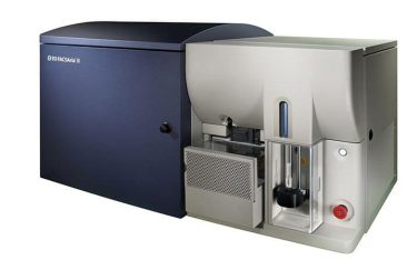
A digital high-speed flow cytometry based cell sorter. Equipped with five lasers line; 375nm, 405nm, 488nm, 561nm and 640nm. the BD FACSAria III Cell Sorter is designed to precisely sort cells at high flow speeds and to enable simultaneous multicolor analysis of up to 18 parameters. Sorted cells (up to four distinct populations) are collected in a variety of collecting tubes or multi-well plates, with three sizes of nozzles (70, 85 and 100 micron). There is a temperature controlled sample chamber and collection tube holder to enhance cell viability.
Sorted cells (up to four distinct populations) are collected in a variety of collecting tubes or multi-well plates, with four sizes of nozzles (70, 85, 100 and 130 micron). There is a temperature controlled sample chamber and collection tube holder to enhance cell viability.
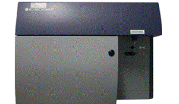
High-performance multicolor flow cytometry analysis. Configured with three lasers; Violet 405, Blue 488 and Red 640, the BD FACSCanto™ II Cell Analyzer delivers high-performance multicolor analysis.
The instrument can accommodate the detection of up to 7 colors simultaneously. Its optical design maximizes signal detection and increases sensitivity and resolution for each color in a multicolor assay.
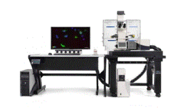
Confocal laser scanning microscope and fluorescent microscope for advanced imaging Configured with three lasers; Violet 405, Blue 488 and Red 640, the BD FACSCanto™ II Cell Analyzer delivers high-performance multicolor analysis.
The instrument can accommodate the detection of up to 7 colors simultaneously. Its optical design maximizes signal detection and increases sensitivity and resolution for each color in a multicolor assay.
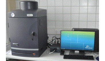
Market trusted technology offering the fullest suite of leading imaging technologies, reagents and support Exquisite sensitivity in bioluminescence Full fluorescence tunability through the NIR spectrum Compute Pure Spectrum spectral unmixing for ultimate fluorescence sensitivity
Preclinical imaging for non-invasive longitudinal monitoring of disease progression, cell trafficking and gene expression patterns in living animals.
The IVIS Lumina III is capable of imaging both fluorescent and bioluminescent reporters. The system is equipped with up to 26 filter sets that can be used to image reporters that emit from green to near-infrared. Superior spectral unmixing is achieved by Lumina III’s optional high resolution short cut off filters.
Features & Benefits
- Market trusted technology offering the fullest suite of leading imaging technologies, reagents and support
- Exquisite sensitivity in bioluminescence
- Full fluorescence tunability through the NIR spectrum
- Compute Pure Spectrum spectral unmixing for ultimate fluorescence sensitivity
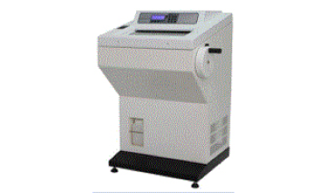
Produces high-quality tissue sections for in situ staining. The cryostat preserves frozen tissue samples in low cryogenic temperatures, while a microtome slices the tissue into pieces thin enough to be observed under a microscope.
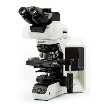
INCLUDES:
3 POSITION TRINOCULAR HEAD (100% EYES/ 100%CAMERA/ SPLIT BETWEEN EYES & CAMERA)
10X WIDEFIELD EYEPIECES
UPLANFL 4X OBJECTIVE<br>
UPLANFL 10X OBJECTIVE<br>
UPLANFL 20X OBJECTIVE<br>
UPLANFL 40X OBJECTIVE<br>
UPLANFL 100X OIL IMMERSION OBJECTIVE.<br>
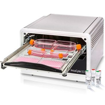
Derive deeper and more physiologically relevant information about your cells – plus real-time kinetic data. The Incucyte® automatically acquires and analyzes images around the clock, providing an information-rich analysis that is easy to achieve.
Change can happen in an instant. Whether simply assaying cell health or more complex processes like migration, invasion, or immune cell killing, see what happened and when—without ever removing your cells from the incubator.<br>
Never miss powerful insights again, with the Incucyte® S3 live-cell analysis system, reagents, and consumables.
